bone anatomy ankle
Ankle bones, talus, navicular, cuneiforms, calcaneus, and cuboid. 9 Pics about Ankle bones, talus, navicular, cuneiforms, calcaneus, and cuboid : Muscle Bone Attachments, Ankle bones, talus, navicular, cuneiforms, calcaneus, and cuboid and also Normal ankle joint, frontal X-ray. — front view, human body - Stock.
Ankle Bones, Talus, Navicular, Cuneiforms, Calcaneus, And Cuboid
 www.pinterest.com
talus appendicular navicular cuboid calcaneus cheville cuneiforms tarsus tarsal naviculaire tarse cuboïde visiblebody squelette anatomie consists
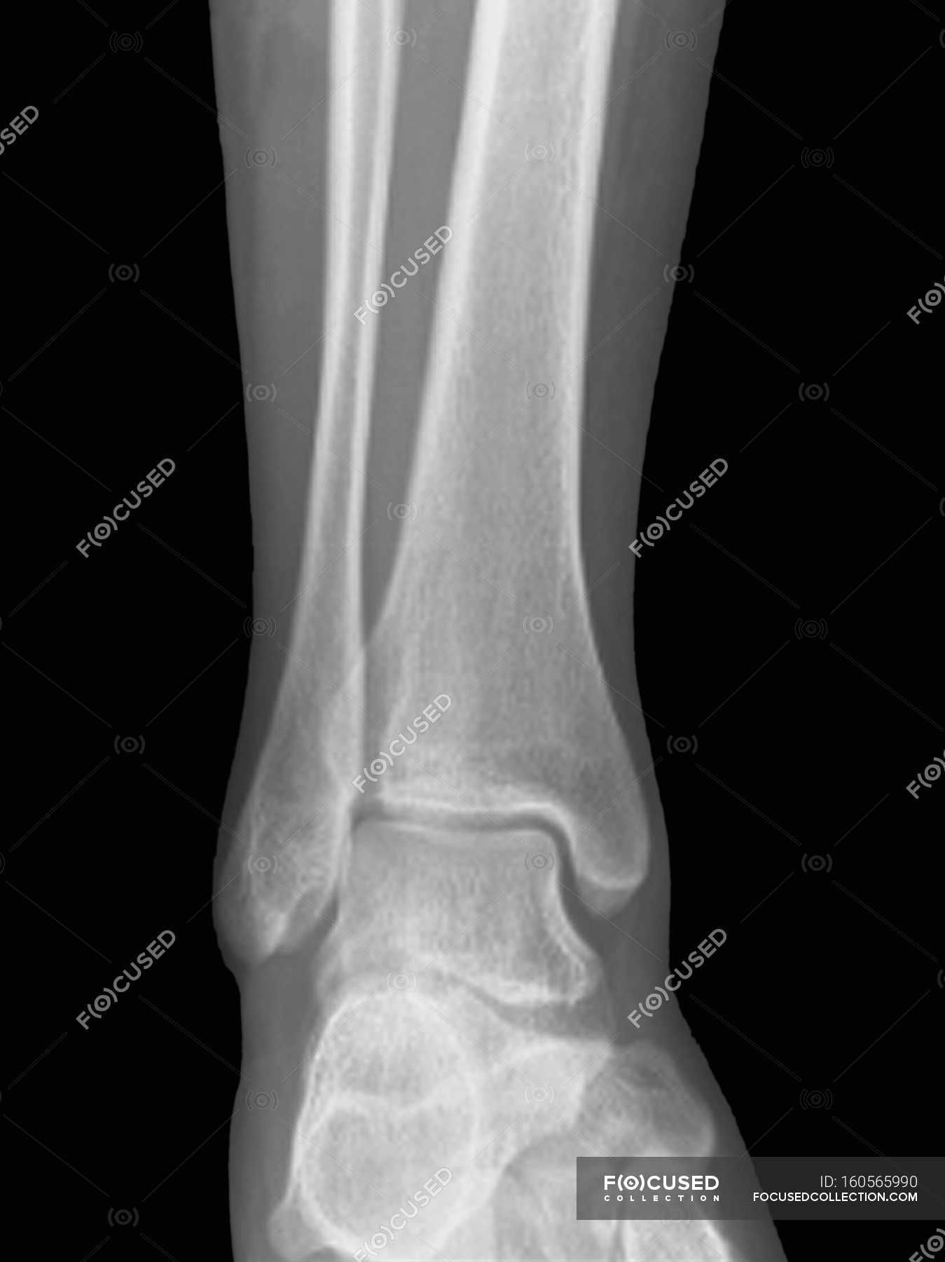
Normal Ankle Joint, X-ray - Stock Image - F002/7554 - Science Photo Library
 www.sciencephoto.com
ankle normal ray joint
The Hip Joint - Complete Physiotherapy
 www.completephysiotherapy.co.uk
hip joint anatomy human bones body replacement ball resurfacing bone pelvis femur skeletal where system virtual diagram ligament spine conditions
Normal Ankle Joint, Frontal X-ray. — Front View, Human Body - Stock
 focusedcollection.com
ankle frontal
Avascular Necrosis Of Navicular Bone | Image | Radiopaedia.org
 radiopaedia.org
navicular bone necrosis avascular radiopaedia sagittal t1
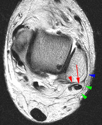
Peroneal Tendon Dislocation And Superior Peroneal Retinaculum Injury
 radsource.us
peroneus anatomy mri peroneal tendon retinaculum brevis section normal dislocation quartus 4b superior injury radsource 2006 muscle skeletal musculo
Muscle Bone Attachments
 www.anatomyfacts.com
muscle attachments bone anatomy ankle foot plantar posterior anterior feet forearm b3 e3 body
Stress Fracture | Image | Radiopaedia.org
 radiopaedia.org
fracture stress radiopaedia cortical radiology fibrous defect version frontal
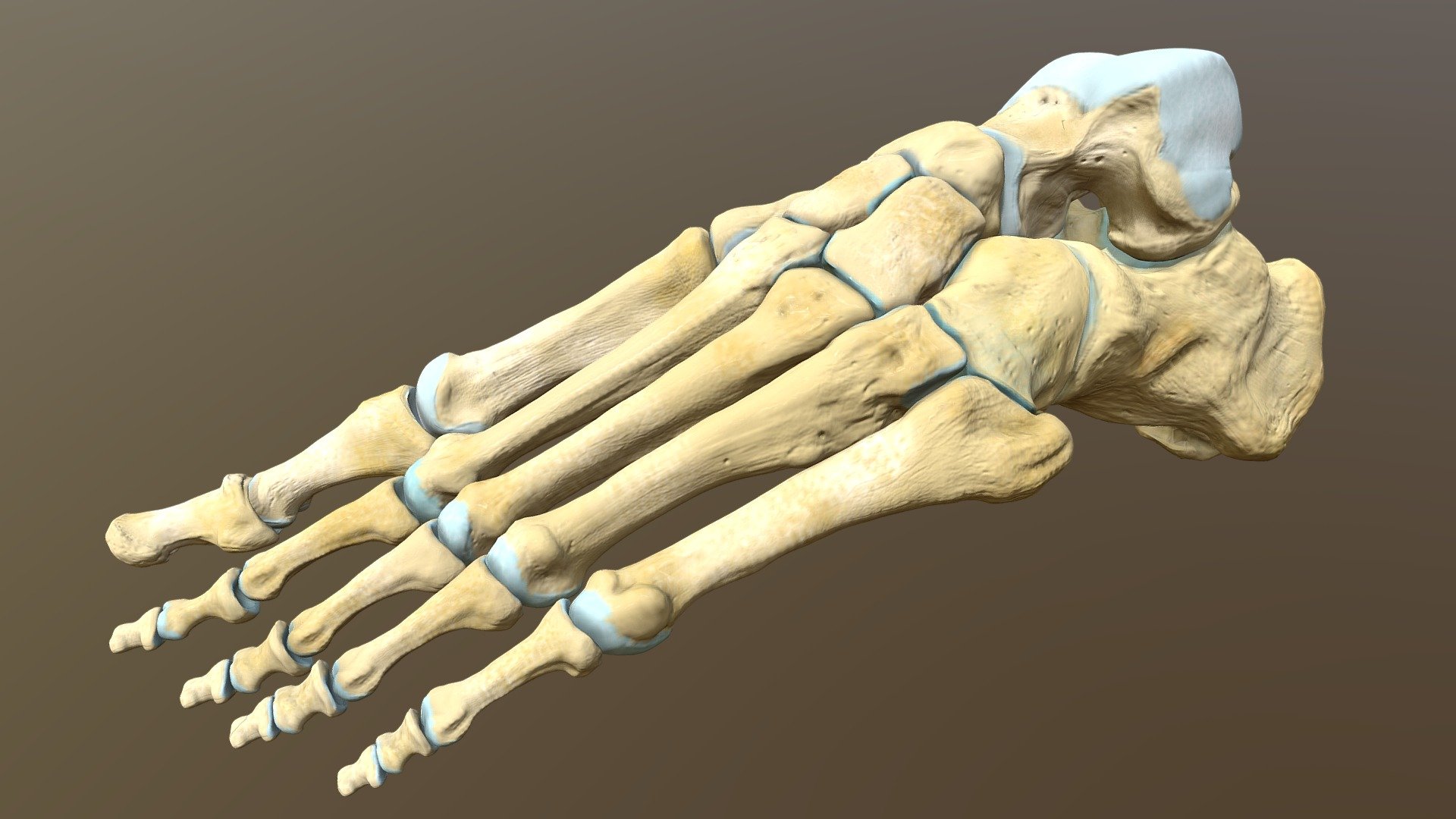
Left Human Foot Bones - Buy Royalty Free 3D Model By Catherine Sulzmann
 sketchfab.com
sketchfab sulzmann
Left human foot bones. Peroneal tendon dislocation and superior peroneal retinaculum injury. Sketchfab sulzmann