c1 bone anatomy
Interpreting cervical spine radiographs | The BMJ. 9 Pictures about Interpreting cervical spine radiographs | The BMJ : Lateral View of Skull and Cervical Spine | Neuroanatomy | The, Medical Pictures Info – Cervical Vertebrae and also Posterior View of Spinal Cord at Level of Foramen Magnum | Neuroanatomy.
Interpreting Cervical Spine Radiographs | The BMJ
 www.bmj.com
cervical bmj radiographs interpreting odontoid
Lateral Plain Radiograph Of The Cervical Spine | The BMJ
 www.bmj.com
lateral spine cervical radiograph plain bmj
Medical Pictures Info – Cervical Vertebrae
 medicalpicturesinfo.com
c7 cervical vertebrae spine neck vertebra disc pain herniated spinal c1 symptoms nerve anatomy ray where c5 levels vertebral c2
Atlanto Occipital Joint Is A Common Cause Of Headache Which
 www.chiropractic-help.com
occipital atlanto instability consequences misalignment
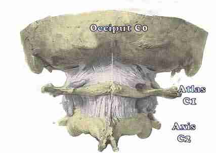
Transoral View Of Skeletal Anatomy | Neuroanatomy | The Neurosurgical
 www.neurosurgicalatlas.com
skeletal transoral neurosurgicalatlas correlation
Lateral View Of Skull And Cervical Spine | Neuroanatomy | The
 www.neurosurgicalatlas.com
spine lateral neuroanatomy
Levator Scapulae Muscle
 www.yoganatomy.com
levator scapulae scapula muscle
Posterior View Of Spinal Cord At Level Of Foramen Magnum | Neuroanatomy
 www.neurosurgicalatlas.com
spinal foramen cord magnum posterior level atlas surgical correlation neurosurgicalatlas
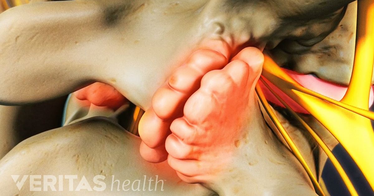
Cervical Osteophytes: Bone Spurs In The Neck
 www.spine-health.com
bone spurs cervical neck osteophytes spine pain osteoarthritis lumbar arthritis facet spondylosis disc treatment spur shoulder called symptoms spinal causes
Levator scapulae muscle. Lateral plain radiograph of the cervical spine. Occipital atlanto instability consequences misalignment